Happy to be stuck with you
| 13 January, 2011 | Richard P. Grant |
|
|
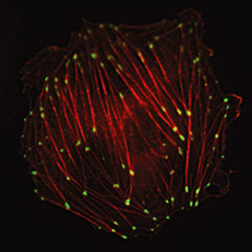
Talin and actin
We’ve not had any decent cell biology here for a while, but there’s a rather nice controversy that boiled up last year about the presence—or otherwise—of adherens junctions—focal contacts—in 3D cell cultures. A paper from Denis Wirtz’s lab made the rather striking claim that live cells migrating through a (more physiological) three-dimensional matrix do not form the focal adhesions that characterize cells in 2D culture. The usual suspects of focal adhesions—vinculin, paxillin, talin, alpha-actinin, zyxin, VASP, focal adhesion kinase—were present, but distributed diffusely throughout the cytoplasm.
The paper attracted no fewer than seven evaluations and a comment, giving it an F1000 Factor of 17. Clearly, were these results to be replicated it would be a very interesting and important finding indeed.
But at the meeting of the American Society for Cell Biology in December, we heard rumours of a paper in press refuting this finding. And indeed, just before Christmas came the news (again in Nature Cell Biology) that reducing background fluorescence reveals adhesions in 3D matrices. Fraley et al. have responded, although Faculty Member Ken Yamada remains unconvinced by their arguments in his evaluation.
Ken, who I interviewed early last year for a The Scientist article on a novel and relatively easy way of looking at three dimensional growth in culture, admits there is a problem for us mortals:
Consequently, readers might seem to be left with a dilemma, because these two excellent research teams present contradictory findings using the same type of 3D collagen gel and the same cell lines.
He does, however, come out pretty strongly on the side of Kubow and Horwitz, saying
If one research group sees and quantifies a structure and another group reports that they cannot see it, the structure presumably exists unless it can be shown to be an experimental artifact – which does not appear to be the case here. It will be interesting to learn later what experimental problems encountered by Fraley et al. limited their visibility of 3D adhesions, which may help others perform these types of study in the future.
Reinhard Fässler and Herbert Schiller have also independently recommended the Kubow and Horwitz paper as an F1000 must read.
So what happens now? According to Ken Yamada, the field can now proceed to characterize these structures for their tissue-specificity and functions . And breathe a sigh of relief, presumably.
|
|
I like the tag for this article, ‘cell porn’
Since the organization of focal contacts reflects the type of substrate that the cell is adhering to, it is not surprising that cells adhering to plastic or glass form adhesions that fundamentally differ from those formed by cells grown inside a soft matrix. The visualization of focal contact organization in cells grown inside a matrix has been (and apparently) still is technically very challenging. An important issue to remember however is that when key focal adhesion components (vinculin, a-actinin) were specifically overexpressed, or their level was suppressed (in studies published almost 20 years ago), this had a dramatic effect on cancer cell growth, motility and tumorigenic capacity in mice.
Thanks for your comment, Avri. But isn’t the point not simply that the adhesions were fundamentally different (I agree: that wouldn’t be surprising), but that they weren’t seen at all? And that it’s more likely that because they’re different, it’s easier to miss them?